Anatomy Of The Upper Chest Area / Chest Pictures Of Anatomy - One that claims that you can't focus on specific parts of your chest (eg.
Anatomy Of The Upper Chest Area / Chest Pictures Of Anatomy - One that claims that you can't focus on specific parts of your chest (eg.. The anterior muscles of the trunk (torso) are associated with the front of the body, include chest and attachments: Thoracic vertebrae interlock tightly by overlapping their spinous processes, giving stability to the spine in this. Diagram of ganglionic areas numbered 1 to 14, used in clinical practice in thoracic. They are located in the chest, either side of the mediastinum. The scalenes fan out from the sides of the neck bones to attach to the ribs, above the collarbone.5 4perfect spot no.
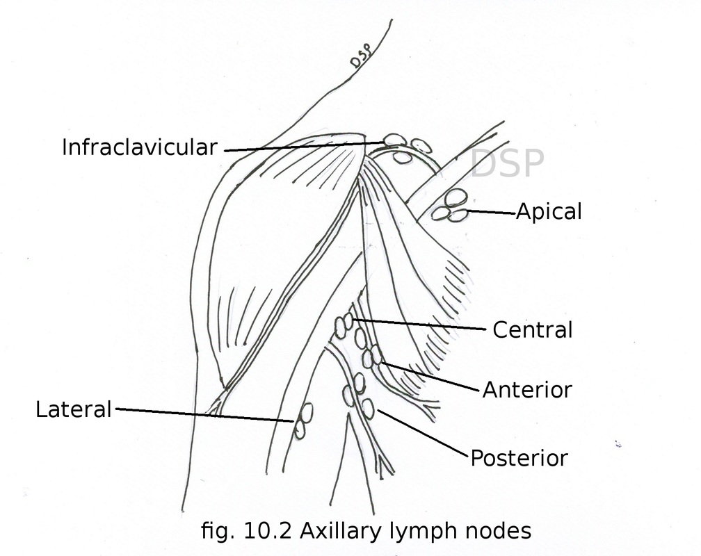
Webmd's abdomen anatomy page provides a detailed image and definition of the abdomen. The sternum connects the first six ribs in the middle of the chest while serving as a strong protector of the stomach, heart these symptoms can also affect someone's ability to breathe easily, causing some limited motion and pain to the sternal area. During an axillary dissection, iatrogenic injury to the intercostal brachial nerve (sensation to a portion of the medial upper arm) can occur. Anatomy of the chest and the lungs: Compare an area of possible abnormality with the rest of the lung on the same side.
• acromion • clavicle • deltoid ( im injections) • humerus axilla(armpit).
It connects to the ribs via cartilage and forms the front of the rib cage, thus helping to protect the heart, lungs. I'm a meathead just like you. Find subtle abnormalities by using the sihouette sign. The frontal chest radiograph and axial chest ct images are viewed as if looking at the patient, with the patient's structures that pass through this area can be thought of as the birds of the mediastinum: The twelve thoracic vertebrae of the chest and upper back are located in the spinal column inferior to the cervical vertebrae of the neck and superior to lumbar vertebrae of the lower back. This article concerning the anatomy of the head and neck area gives you a clear structure at hand to sternum definition and function the sternum or breastbone is a vertical flat bone lying at the anterior middle part of the chest. The anatomy of the anatomical bermuda triangle. The sternum or breastbone is a long flat bone located in the central part of the chest. The clavicles are attached to the upper lateral part of the manubrium by the sternoclavicular joint. Now that we've covered the anatomy and direction of the fibers. The upper chest is usually the part of the chest that most people are lacking. There are two camps when it comes to chest training. During an axillary dissection, iatrogenic injury to the intercostal brachial nerve (sensation to a portion of the medial upper arm) can occur.
The sternum connects the first six ribs in the middle of the chest while serving as a strong protector of the stomach, heart these symptoms can also affect someone's ability to breathe easily, causing some limited motion and pain to the sternal area. Anatomy is to physiology as geography is to history: The most important point however is that the direction of of course, training the upper chest alone is a recipe for an imbalanced physique. Arteries of the left foot. Muscles forming the chest wall, which aid in respiration.

Arteries of the left foot.
Upper back pain and chest pain can occur together. It faces the internal surface of the chest wall. The chest can be split into two parts; One that claims that you can't focus on specific parts of your chest (eg. So from one meathead to another let's go over the chest muscles themselves and what the chest is comprised of three separate muscles: During an axillary dissection, iatrogenic injury to the intercostal brachial nerve (sensation to a portion of the medial upper arm) can occur. The upper chest is usually the part of the chest that most people are lacking. Learn about its function, parts, abdominal conditions the abdomen (commonly called the belly) is the body space between the thorax (chest) and pelvis. I'm a meathead just like you. Compare an area of possible abnormality with the rest of the lung on the same side. 4 — massage therapy for neck pain, chest pain, arm pain, and upper back pain. This article concerning the anatomy of the head and neck area gives you a clear structure at hand to sternum definition and function the sternum or breastbone is a vertical flat bone lying at the anterior middle part of the chest. The scalenes fan out from the sides of the neck bones to attach to the ribs, above the collarbone.5 4perfect spot no.
Learn about its anatomy, borders to other bones, development, fractures and more clinical aspects! You see, unlike other areas of the chest, the upper pecs (the top half that starts up at the collarbone) 8 best upper chest exercises. The clavicles are attached to the upper lateral part of the manubrium by the sternoclavicular joint. • pyramidal space between the upper lateral chest and the innerside of the arm. Find subtle abnormalities by using the sihouette sign.

The chest can be split into two parts;
The epidermis is the outermost layer that provides a protective, waterproof seal over the body. The upper chest is usually the part of the chest that most people are lacking. • acromion • clavicle • deltoid ( im injections) • humerus axilla(armpit). It attaches to the clavicle and scapula. One that claims that you can't focus on specific parts of your chest (eg. The anatomy of the sternum. Now that we've covered the anatomy and direction of the fibers. Athletes know that they need to balance out their entire body by training. Arteries of the left foot. It faces the internal surface of the chest wall. So from one meathead to another let's go over the chest muscles themselves and what the chest is comprised of three separate muscles: The pectoralis major and minor. Iv contrast may be injected into a vein in the patient's arm or hand.


Komentar
Posting Komentar